From biological event to
measurable evidence.
Preserve structures in blood, identify targets by their molecular markers, and measure their abundance and morphology.
How the platform works.
From preserved blood to structure-resolved NETosis data.
- Preserve the sample
- Label selected markers
- Image the population
- Measure individual events
One sample.
Evidence at every scale.
Explore a whole-blood sample, then select an event to compare DNA, H3Cit and MPO signals within the same structure.
Inspect the complete gel stripe.
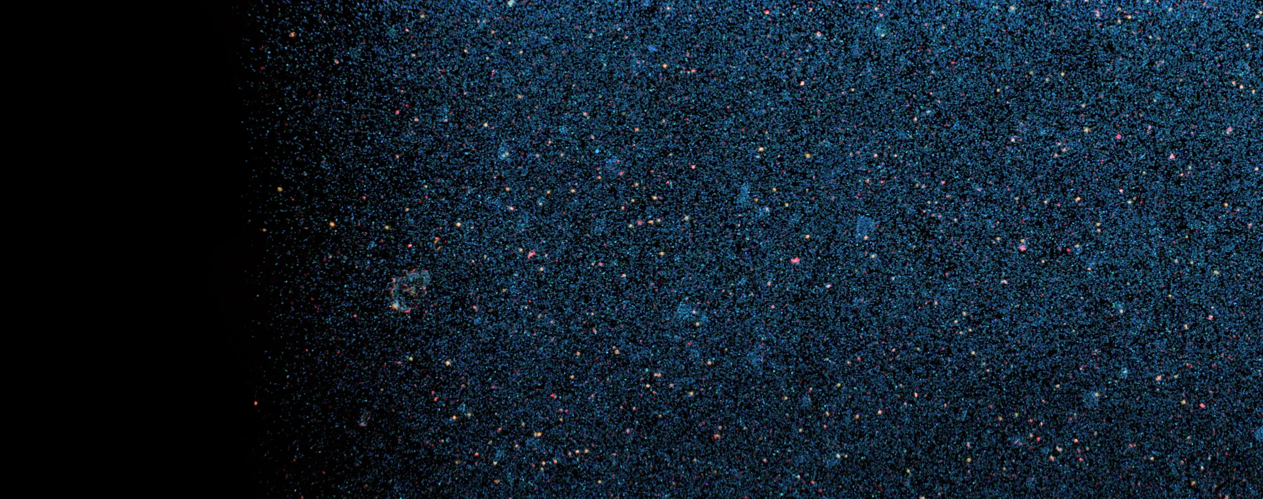



ZOOM
Event Gallery.
Select any H3Cit-associated event to inspect its DAPI, SYTOX, H3Cit and MPO channels.
Compare these events with curated cellular phenotypes and larger extracellular structures.
Explore the NETosis AtlasFrom events to population profiles.
Generated example data show how event counts, marker signals and structure size contribute to a sample profile.
Sample signature
The higher-burden example relative to the lower-burden example.
Compare the populations
Both examples use the same assumed sample size and axes.
Lower-burden example
5,000 H3Cit-selected targets.
Higher-burden example
9,000 H3Cit-selected targets.
ONE H3Cit-SELECTED TARGETMPO COLOCALIZATION · LOW / MID / HIGHEVENT AREA
ReadoutLower burdenHigher burden
H3Cit+ targets5,0009,000
MPO-confirmed70.7%70.3%
Qualified events3,5366,323
Mean event area1.0×1.9×
DNA expansion1.0×1.9×
Large-event fraction1.3%30.1%
Integrated burden1.0×3.4×
How the readouts are calculated
In these generated examples, qualified events have mid/high MPO colocalization. Integrated burden sums their normalized event areas; large events have normalized area ≥ 0.50.
DNA expansion, mean area and integrated burden are shown relative to the lower-burden example. Both populations assume the same sample size.
Panels built around
the biology.
Extend the marker panel beyond DNA, H3Cit and MPO to investigate cell lineage, pathway state, molecular cargo or cell–NET interactions. Targets and interpretation criteria are defined for each study.
Registered images
Whole-sample views and multichannel images of individual events, with measurements traceable to the source images.
Quality checks and measurements
Processing quality checks and study-defined readouts of event abundance, morphology, marker localization and interactions.
Research report
Methods, results and interpretation limits brought together with the agreed endpoint tables.